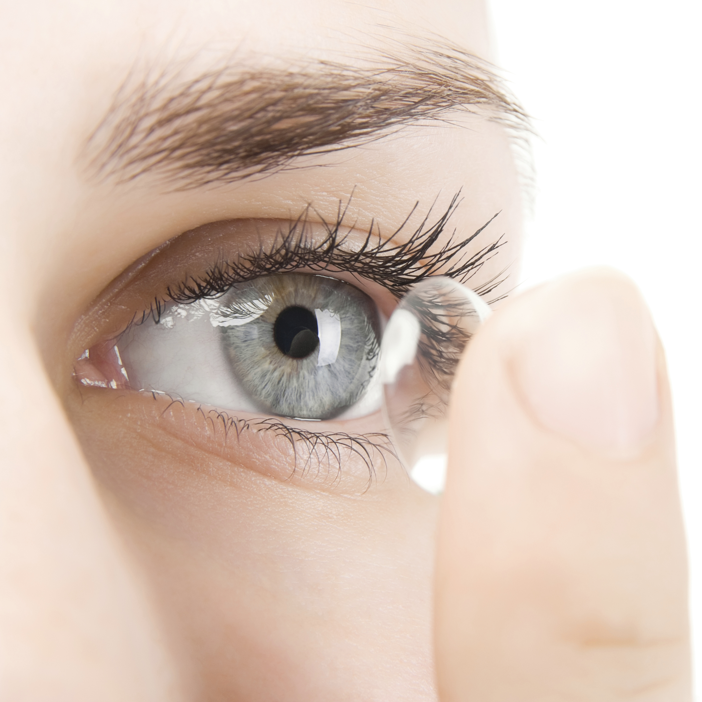

5 A contact lens with a biosensor may be useful for monitoring both rate and completeness of an individual’s blink. It is well known that blink rate is reduced during near activities, such as computer use and reading. Another limitation is the indirect IOP measurement by corneal curvature compared with in-office applanation tonometry measurements, potentially making it difficult for the clinician to compare and determine next steps for clinical management.Ĭontact lenses may also aid in the diagnosis of dry eye disease by measuring tear film osmolarity, inflammatory cytokines, and blink rate. One limitation of this lens is potential discomfort due to its thickness (two to three times that of a traditional soft lens, and/or its stiff modulus). The most common side effects are transient blur, conjunctival hyperemia, and punctate keratitis, which have been reported to resolve within 24 to 48 hours. The device provides 288 IOP data points throughout a 24-hour period, is available in three base curves, and has an oxygen transmissibility of 119 Dk/t. 4 It uses an 11.5 mm–wide circular gauge that corresponds to the corneo-scleral junction and location of highest deformation due to IOP changes. The Sensimed Triggerfish (Sensimed) continuous ocular monitoring system is FDA approved for the 24-hour measurement of IOP. An IOP change of 1 mm Hg produces a 3 mm change in corneal curvature in an average-sized cornea, and devices have been developed to detect such changes. Contact lens technology can provide accurate readings over a length of time.

In-office IOP readings are limited because they provide only a static IOP reading (ie, at one point in time). IOP readings, specifically dynamic measurements that account for circadian changes and short-term fluctuations, are critical in the management of patients with glaucoma, especially those with end-stage or progressive disease. Contact lenses may be useful in acquiring direct measurement with biosensors or indirect measurement of accumulation of tear properties on the lens and after removal. A tear film protein called lacryglobin is elevated in patients with lung, breast, colon, and prostate cancer, and in those with a family history of cancer. This time period can be critical for patients who experience a severe hypo- or hyperglycemic crisis.Ĭancer detection by tear film biomarkers is another area of research in the contact lens space. The main drawback in glucose monitoring via tear film is the 20-minute lag between changes in blood glucose levels and changes in tear glucose levels. The electrochemical method is more accurate, although more cumbersome compared with the optical detection method. The electrochemical method relies on more complex components and chemical reactions to produce an electrical current proportional to glucose within the tear film.

3 Optical detection relies on glucose binding to sensors, resulting in fluorescent change and interpretation by a photodetector or smartphone. Two potential noninvasive means of glucose monitoring using contact lenses are being researched: the optical detection method 2 and the electrochemical method. Blood glucose is typically monitored by the finger-prick method, a procedure that uses a lancet to acquire a blood sample. The tear film of a patient with diabetes will have elevated glucose, advanced glycation end proteins, and cytokine changes. For example, Alzheimer disease presents with increased levels of dermcidin, lacritin, lipocalin-1, and lysozyme-C in the patient’s tear film. The tear film was previously thought to be a biomarker only for ocular disease detection, but its immune properties can also assist in diagnosing various systemic conditions.


 0 kommentar(er)
